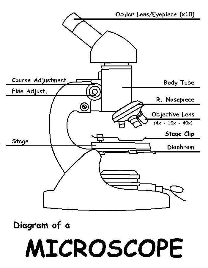
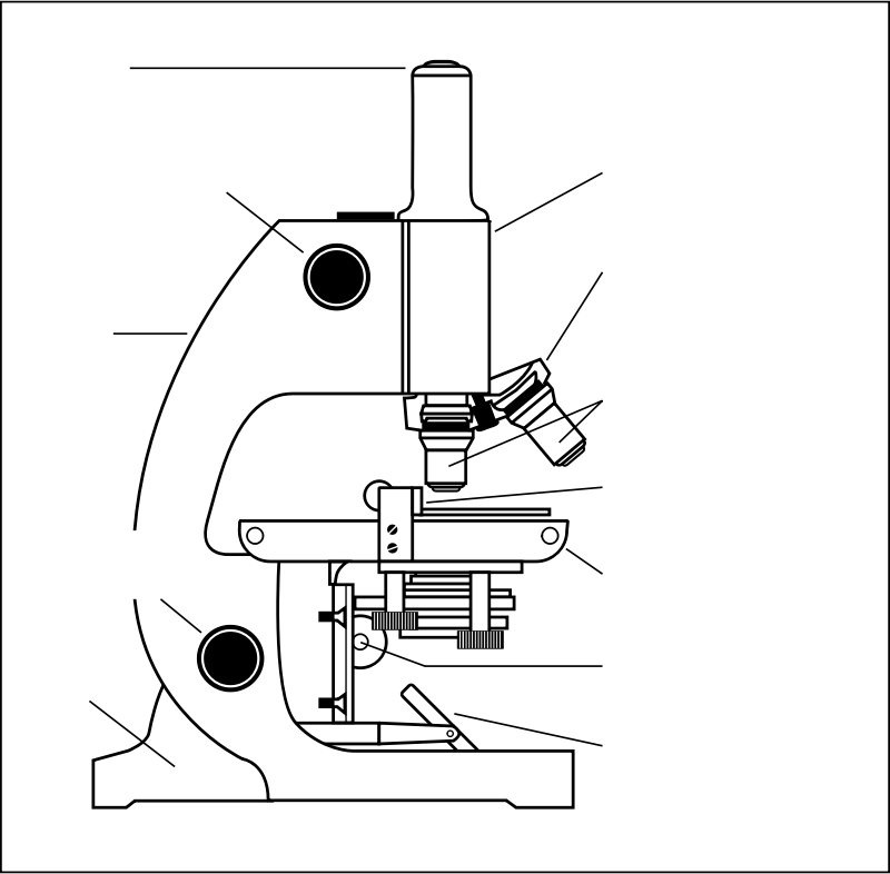
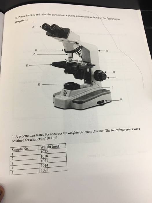
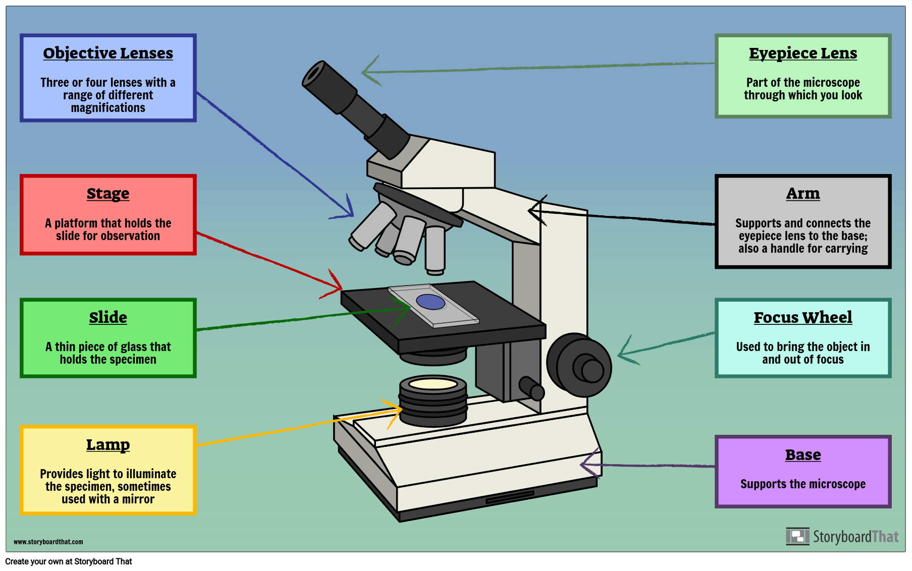
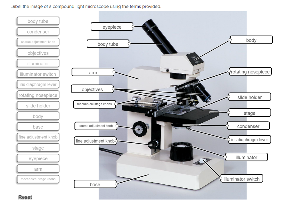
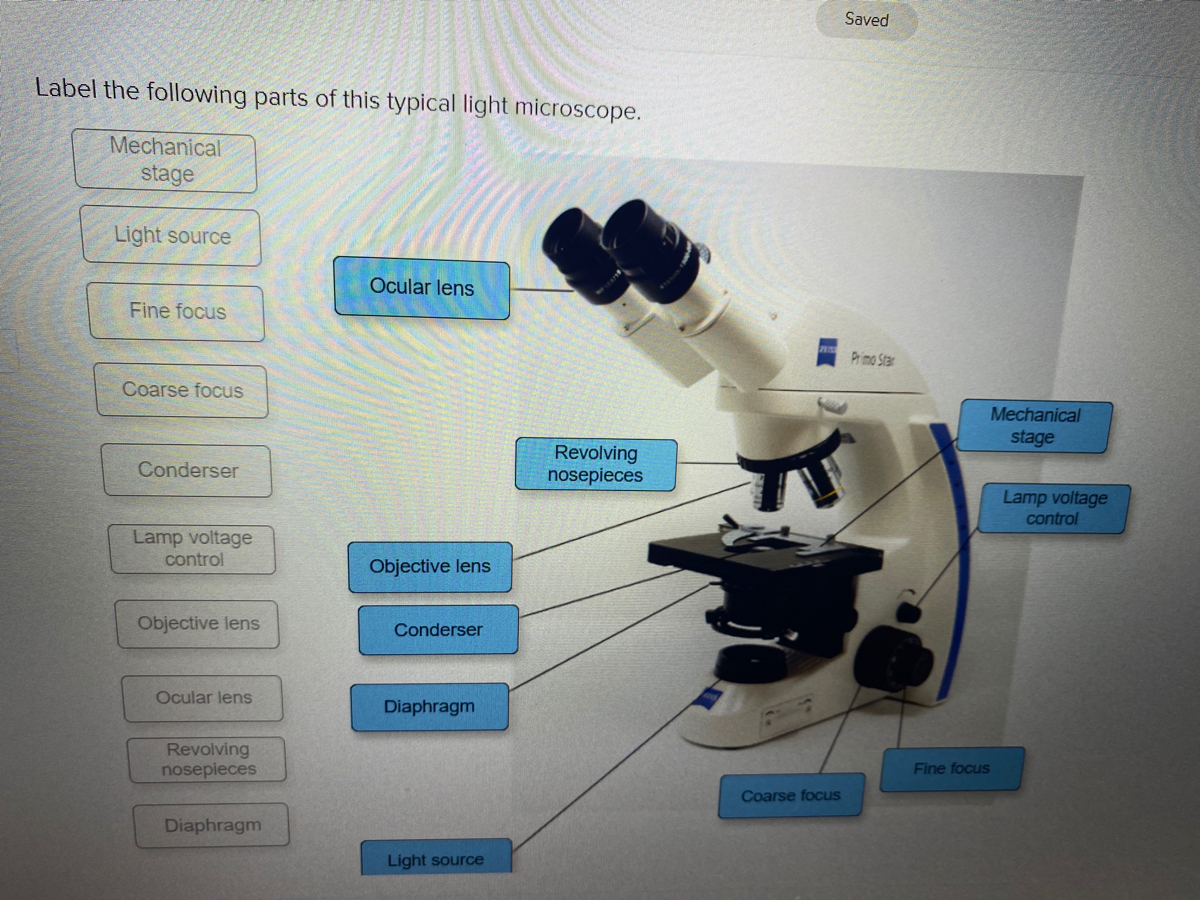
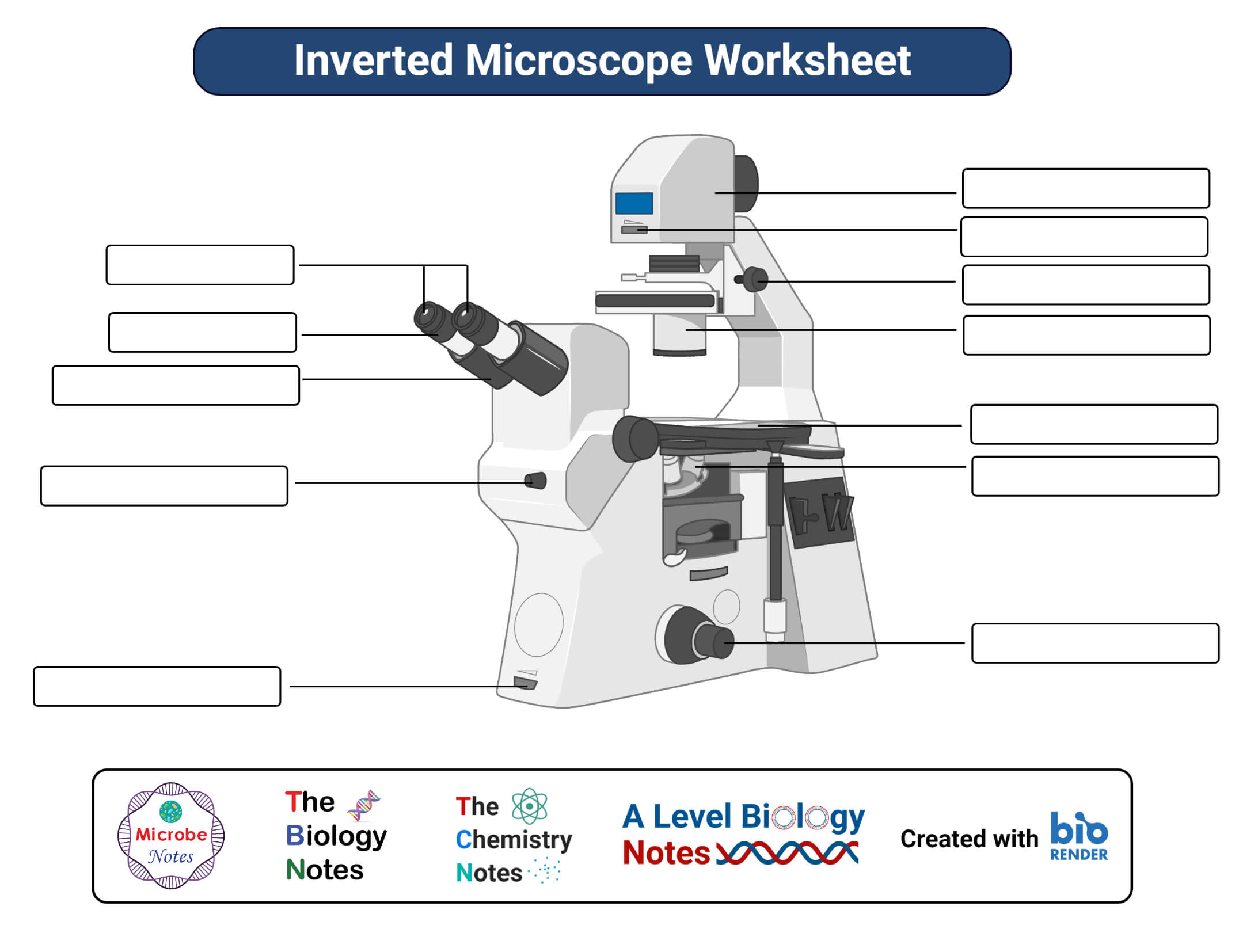
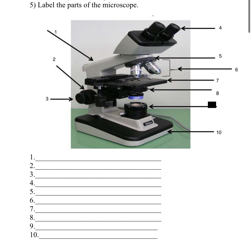
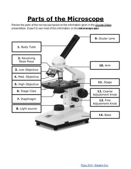
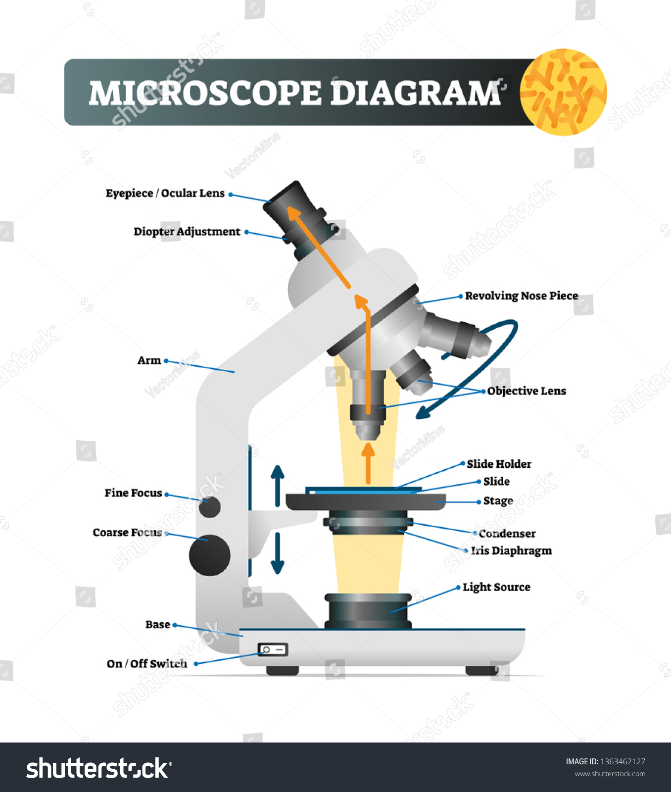
38 picture of microscope with label
PDF Parts of a Microscope Printables - Homeschool Creations Label the parts of the microscope. You can use the word bank below to fill in the blanks or cut and paste the words at the bottom. Microscope Created by Jolanthe @ HomeschoolCreations.net. Parts of a eyepiece arm stageclips nosepiece focusing knobs illuminator stage objective lenses PDF Label parts of the Microscope Label parts of the Microscope: . Created Date: 20150715115425Z
Compound Microscope Labeled Diagram | Quizlet QUESTION. The total magnification of a specimen being viewed with a 10X ocular lens and a 40X objective lens is. 15 answers. QUESTION. a mosquito beats its wings up and down 600 times per second, which you hear as a very annoying 600 Hz sound. if the air outside is 20 C, how far would a sound wave travel between wing beats. 2 answers.
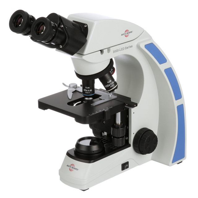
Picture of microscope with label
The Parts of a Microscope (Labeled) Printable - TeacherVision The Parts of a Microscope (Labeled) Printable Download Add to Favorites Share This diagram labels and explains the function of each part of a microscope. Use this printable as a handout or transparency to help prepare students for working with laboratory equipment. Grade: 9 | 10 | 11 | 12 Subjects: Science Scientific Method www1.udel.edu › biology › ketchamUD Virtual Compound Microscope - University of Delaware ©University of Delaware. This work is licensed under a Creative Commons Attribution-NonCommercial-NoDerivs 2.5 License.Creative Commons Attribution-NonCommercial-NoDerivs 2 Parts of a Simple Microscope - Labeled (with diagrams) image 1: The images above are all examples of a simple microscope. image source: laboratoryinfo.com image 2: A simple microscope commonly used by students for studying minute objects. image source: imimg.com picture 3: It is the latest design of a simple microscope - advanced features than the conventional simple microscopes.
Picture of microscope with label. A Study of the Microscope and its Functions With a Labeled Diagram ... The camera present within the microscope captures images to reveal the finer details of the specimen. This microscope can zoom and view the density of a specimen until it is only a micrometer thick and has a magnification ranging between 1,000 - 250,000x on the fluorescent screen. This microscope needs a computer software to yield precise ... Drawing Of A Microscope And Label - Warehouse of Ideas Yet even with the technology to digital capture images, many scientists still depend on their abilities to sketch microscope slides. Drawn as seen through 400x magnification). Here presented 54+ microscope drawing and label images for free to download, print or share. Title Is Informative, Centered, And Larger Than Other Text. learn.genetics.utah.edu › content › cellsCell Size and Scale - University of Utah Smaller cells are easily visible under a light microscope. It's even possible to make out structures within the cell, such as the nucleus, mitochondria and chloroplasts. Light microscopes use a system of lenses to magnify an image. The power of a light microscope is limited by the wavelength of visible light, which is about 500 nm. Label the microscope — Science Learning Hub Label the microscope Interactive Add to collection Use this interactive to identify and label the main parts of a microscope. Drag and drop the text labels onto the microscope diagram. coarse focus adjustment diaphragm or iris stage eye piece lens fine focus adjustment base high-power objective light source Download Exercise Tweet
scienceexplorers.com › guide-to-teaching-kidsGuide to Teaching Kids About Cells | Science Explorers Apr 25, 2019 · Give each student a chance to look through the microscope at the cells. Point out that each slide contains numerous cells. Repeat the process with the second slide. Have students draw pictures of what they saw under the microscope and guess what the cells do. Finish with an explanation of the cell and its organelle functions. Microscope Labeled Pictures, Images and Stock Photos Browse 49 microscope labeled stock photos and images available, or start a new search to explore more stock photos and images. Newest results Fluorescent Imaging immunofluorescence of cancer cells growing... Microscope diagram vector illustration. Labeled zoom instrument... Microscope diagram vector illustration. › int › scientificXGT-9000 - HORIBA Micro-XRF can be used to understand the elemental distributions in insects non-destructively. In this application note, we carried out elemental map imaging on edible crickets using a HORIBA XGT-9000 X-ray analytical microscope and revealed the rich source of zinc in their jaws. Compound Microscope Parts - Labeled Diagram and their Functions The eyepiece (or ocular lens) is the lens part at the top of a microscope that the viewer looks through. The standard eyepiece has a magnification of 10x. You may exchange with an optional eyepiece ranging from 5x - 30x. [In this figure] The structure inside an eyepiece. The current design of the eyepiece is no longer a single convex lens.
Microscope With Labels clip art | Microscope parts, Scientific method ... vector clip art online, royalty free & public domain Download Clker's Microscope With Labels clip art and related images now. Multiple sizes and related images are all free on Clker.com. D Dixie Tsutsaeva 2k followers More information Microscope With Labels clip art Find this Pin and more on Art Journal Inspiration by Dixie Tsutsaeva. Compound Microscope - Diagram (Parts labelled), Principle and Uses What are the 13 parts of a microscope? 1. Eyepiece 2. Eyepiece Tube 3. Objective Lens 4. Stage 5. Stage Clips 6. Nosepiece 7. Fine and Coarse Focus knobs 8. Illuminator 9. Aperture 10. Iris Diaphragm 11. Condenser 12. Condenser Focus Knob 13. The Rack stop Q 5. What are the 11 parts of a compound microscope? biomedx.com › microscopesMicroscopes | Biomedx Microscope System Limited Warranty Biomedx warrants that the microscope systems it sells and any related accessories bearing the Biomedx label (individually a “Product” and collectively the “Products”) will be free from defects in materials and workmanship under normal use and service for a period, beginning from the date of purchase, of ; Microscope Parts, Function, & Labeled Diagram - slidingmotion Microscope parts labeled diagram gives us all the information about its parts and their position in the microscope. Microscope Parts Labeled Diagram The principle of the Microscope gives you an exact reason to use it. It works on the 3 principles. Magnification Resolving Power Numerical Aperture. Parts of Microscope Head Base Arm Eyepiece Lens
Microscope Parts and Functions First, the purpose of a microscope is to magnify a small object or to magnify the fine details of a larger object in order to examine minute specimens that cannot be seen by the naked eye. Here are the important compound microscope parts... Eyepiece: The lens the viewer looks through to see the specimen.
Parts of the Microscope with Labeling (also Free Printouts) Parts of the Microscope with Labeling (also Free Printouts) By Editorial Team March 7, 2022 A microscope is one of the invaluable tools in the laboratory setting. It is used to observe things that cannot be seen by the naked eye. Table of Contents 1. Eyepiece 2. Body tube/Head 3. Turret/Nose piece 4. Objective lenses 5. Knobs (fine and coarse) 6.
Microscope Types (with labeled diagrams) and Functions The working principle of a simple microscope is that when a lens is held close to the eye, a virtual, magnified and erect image of a specimen is formed at the least possible distance from which a human eye can discern objects clearly. Simple microscope labeled diagram Simple microscope functions It is used in industrial applications like:
467,408 Microscope Images, Stock Photos & Vectors | Shutterstock Microscope royalty-free images 467,408 microscope stock photos, vectors, and illustrations are available royalty-free. See microscope stock video clips Image type Orientation Color People Artists More Sort by Popular Science College and University Healthcare and Medical Jobs/Professions Biology microscope laboratory scientist medicine lens Next
Microscope, Microscope Parts, Labeled Diagram, and Functions Microscope, Microscope Parts, Labeled Diagram, and Functions What is Microscope? A microscope is a laboratory instrument used to examine objects that are too small to be seen by the naked eye. It is derived from Ancient Greek words and composed of mikrós, "small" and skopeîn,"to look" or "see".
Parts of a microscope with functions and labeled diagram - Microbe Notes Q. List down the 18 parts of a Microscope. 1. Ocular Lens (Eye Piece) 2. Diopter Adjustment 3. Head 4. Nose Piece 5. Objective Lens 6. Arm (Carrying Handle) 7. Mechanical Stage 8. Stage Clip 9. Aperture 10. Diaphragm 11. Condenser 12. Coarse Adjustment 13. Fine Adjustment 14. Illuminator (Light Source) 15. Stage Controls 16. Base 17.
Microscope Labeling Game - PurposeGames.com About this Quiz. This is an online quiz called Microscope Labeling Game. There is a printable worksheet available for download here so you can take the quiz with pen and paper. This quiz has tags. Click on the tags below to find other quizzes on the same subject. Science.
en.wikipedia.org › wiki › MicroscopyMicroscopy - Wikipedia The field of microscopy (optical microscopy) dates back to at least the 17th-century.Earlier microscopes, single lens magnifying glasses with limited magnification, date at least as far back as the wide spread use of lenses in eyeglasses in the 13th century but more advanced compound microscopes first appeared in Europe around 1620 The earliest practitioners of microscopy include Galileo ...
Microscope picture label Flashcards | Quizlet Start studying Microscope picture label. Learn vocabulary, terms, and more with flashcards, games, and other study tools.
Microscope Labeling - The Biology Corner Microscope Labeling. Shannan Muskopf May 31, 2018. This simple worksheet pairs with a lesson on the light microscope, where beginning biology students learn the parts of the light microscope and the steps needed to focus a slide under high power. The labeling worksheet could be used as a quiz or as part of direct instruction where students ...
Simple Microscope - Parts, Functions, Diagram and Labelling Picture 4: The picture is a transmission electron microscope. Image source: ysjournal.com. In this article, we are going to tackle a simple microscope, its parts and functions, and its applications. ... Also see : Labeling the parts of the Microscope. Image source: slidesharecdn.com. Simple microscope Parts and Functions.
Labeling the Parts of the Microscope | Microscope activity, Science ... 7 Fun Microscope Activities for Homeschool Elementary Students. My children get very excited using REAL science equipment! Your student might appreciate this hands-on learning. Microscopes and kits are pricier items to invest in — but can be used by multiple children for many years to come. C.
Microscope Stock Photos, Pictures & Royalty-Free Images - iStock Microscope. Microscope.This royalty free vector illustration features the main icon on both white and black backgrounds. The image is black and white and had the background rendered with the main icon. The illustration is simple yet very conceptual. Coronavirus test line icons.
Label Microscope Diagram - EnchantedLearning.com Using the terms listed below, label the microscope diagram. arm - this attaches the eyepiece and body tube to the base. base - this supports the microscope. body tube - the tube that supports the eyepiece. coarse focus adjustment - a knob that makes large adjustments to the focus. diaphragm - an adjustable opening under the stage, allowing ...
Labeling the Parts of the Microscope | Microscope World Resources Labeling the Parts of the Microscope This activity has been designed for use in homes and schools. Each microscope layout (both blank and the version with answers) are available as PDF downloads. You can view a more in-depth review of each part of the microscope here. Download the Label the Parts of the Microscope PDF printable version here.
300+ Free Microscope & Laboratory Images - Pixabay 399 Free images of Microscope Related Images: laboratory science bacteria research scientist lab biology chemistry medical Find your perfect microscope image. Free pictures to download and use in your next project.
Microscope Labeling - The Biology Corner Students label the parts of the microscope in this photo of a basic laboratory light microscope. Can be used for practice or as a quiz. ... Microscope Labeling . Microscope Use: 15. When focusing a specimen, you should always start with the _____ objective. 16. When using the high power objective, only the _____ knob should be used. 17. The ...
› games › virtuallabsusingthemicroscopeVirtual Labs: Using the Microscope - GameUp - BrainPOP. In this free online science interactive, students learn the procedures for operating a compound optical light microscope as they would use in a science lab. bVX0-zncj9qJ3G1_r18rkIpQL02X-Oi6tWViR4g4-vwDVmU50WZA-4bRZMjM2TXmc88PAkJ1g0jIembnEbM
Parts of a Simple Microscope - Labeled (with diagrams) image 1: The images above are all examples of a simple microscope. image source: laboratoryinfo.com image 2: A simple microscope commonly used by students for studying minute objects. image source: imimg.com picture 3: It is the latest design of a simple microscope - advanced features than the conventional simple microscopes.
www1.udel.edu › biology › ketchamUD Virtual Compound Microscope - University of Delaware ©University of Delaware. This work is licensed under a Creative Commons Attribution-NonCommercial-NoDerivs 2.5 License.Creative Commons Attribution-NonCommercial-NoDerivs 2
The Parts of a Microscope (Labeled) Printable - TeacherVision The Parts of a Microscope (Labeled) Printable Download Add to Favorites Share This diagram labels and explains the function of each part of a microscope. Use this printable as a handout or transparency to help prepare students for working with laboratory equipment. Grade: 9 | 10 | 11 | 12 Subjects: Science Scientific Method
















Post a Comment for "38 picture of microscope with label"