40 correctly label the following anatomical parts of a kidney.
Correctly label the following anatomical parts of a kidney. Start studying Correctly label the following anatomical parts of a kidney.. Learn vocabulary, terms, and more with flashcards, games, and other study tools. Kidney and Nephron Anatomy Quiz - Registered Nurse RN This is a quiz on the anatomy of the kidney and nephron. Before you start studying the renal system for NCLEX, it is very important you understand the basic anatomy and physiology of the kidney and nephron. These structures are affected by disease processes of the renal system and can lead to various signs and symptoms. This quiz and review will start our renal series.
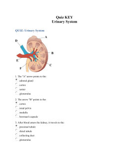
Labeled Diagram of the Human Kidney - Bodytomy Each kidney contains more than 800,000 nephrons, each of which serves as the basic functional unit by performing three essential processes: filtration, reabsorption and secretion. The nephron essentially comprises renal corpuscle and renal tubule. Renal corpuscle

Correctly label the following anatomical parts of a kidney.
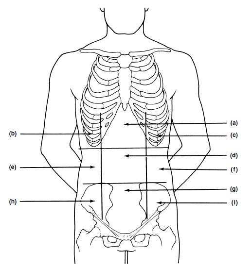
Correctly label the following anatomical features of t… - ITProSpt All of the above. So let's look A. It does indeed consist of vertebrae and inter vertebral cartilage discs. It is reversible. Con B. Is a s trait in humans. It is because we want to keep our head directly above our body for balance, so P. Is also through, and it does enclose the spinal cord to protect it, so the answer is d. The Structure and Function of the Kidneys - Verywell Health What Are the Kidneys? A pair of bean-shaped organs, the kidneys sit in the flanks, closer to the spine than to your belly. They are located just underneath your diaphragm and rib cage. They normally range in size from 8 to 14 centimeters (or 3 to 5.5 inches). Each kidney weighs between 120 grams (about a quarter-pound) to 170 grams (0.4 lbs). PDF BIO 113 LAB 1. Anatomical Terminology, Positions, Planes, and Sections ... Identify and use anatomical terms to correctly label the following regions on Figure 1: BIO 113 Fall 2011 LAB 1 Page 2 ... dividing it into right and left parts, is a sagittal plane. If it divides the body into equal parts, down the midline of the body, it is called a median, or midsagittal, plane. ... kidney and transversely and longitudinally ...
Correctly label the following anatomical parts of a kidney.. To label: The parts of the kidney and nephron. - bartleby The kidney has two zones namely renal cortex and renal medulla. Renal cortex is the outer part of kidney. It consists of blood vessels that are connected to nephrons. Renal medulla is the inner part of the kidney containing 8-12 renal pyramids consisting of collecting tubules. Each kidney has a medially located space called renal sinus. PDF The Urinary System - Pearson 24.2 | Anatomy of the Kidneys 943 Let's now take a closer look at the anatomy of the kidneys. In this module, we explore first the external and internal anatomy of the kidneys. We then turn our attention to the structure and basic roles of the kidneys' functional units: the (NEF-ronz; nephrons nephro- = "kidney"). We conclude the module ... Mastering Chpt 26 - Subjecto.com Which of the following correctly describes the kidneys? The inferior vena cava is medial to the kidneys. The ureters. pass anterior to the iliac artery and vein. Adjacent renal pyramids are separated by the. renal columns. The outer layer of the kidney is the. cortex. Four or five minor calyces join to form. a major calyx. Urinary System Anatomy and Physiology: Study Guide for Nurses The small molecules that result from digestion are absorbed through the walls of the intestine for use in the body. A: This is a function of the Urinary System. The kidneys play an important role in controlling blood levels of Ca 2+ by regulating the synthesis of vitamin D. B: This is a function of the Urinary System.
PDF Anatomy of the Kidney Labeling of the Kidney Overview Health Science Technology students will draw and label the kidney, using proper medical terminology, and will provide a correct definition of the term as follows: Number: Label the structures of the kidney, using the terms provided. Accuracy of Information: Using complete sentences, define each term labeled on ... Name the parts of the nephron - healthsystem.netlify.app This post Parts Of The Nephron Diagram belong to following categorycategories You may also find more related and detailed contents in these categories. Urinary system anatomy Sagittal Section Of The Kidney. The nephron is formed by 4 components corpuscle proximal convoluted tubule Loop of Henle distal convoluted tubule. Solved Correctly label the following anatomical parts of a - Chegg Question: Correctly label the following anatomical parts of a kidney. Ureter Renal pelvis Renal blood vessels Renal column Pyramid of renal modulla Major calyx Renal sinus Renal cortex Renal papilla Minor calyx < Prey 5 of 20 Next > This problem has been solved! See the answer Show transcribed image text Expert Answer 100% (13 ratings) Correctly label the following components of the kidney. Diagram | Quizlet Correctly label the following components of the kidney. STUDY Learn Write Test PLAY Match + − Created by Sarah_Branning Terms in this set (7) Renal artery & vein ... Segmental artery ... Renal cortex ... Renal medulla ... interlobular artery & vein ... Cortical radiate ... Arcuate ...
Correctly label the following anatomical features of a… - ITProSpt Match the key terms with the bone descriptions that follow. Key: (a) clavicle (b) ilium (c) ischium (d) pubis (e) sacrum (f) scapula (g) sternum $-(1)$ bone of the axial skeleton to which the pectoral girdle attaches $-(2)$ markings include glenoid cavity and acromion $-(3)$ features include the ala, crest, and greater sciatic notch $-(4)$ doubly curved; acts as a shoulder strut $-(5)$ hip ... Solved Correctly label the following anatomical parts of a - Chegg Expert Answer 100% (44 ratings) Answer The label is indicated from right … View the full answer Transcribed image text: Correctly label the following anatomical parts of a kidney Renal vein Renal medulia Renal cortex Ureter Renal sinus Renal artery Renal pyramid Renal pelvis Previous question Next question Kidneys: Anatomy, Location, and Function - Verywell Health The kidneys are sandwiched between the diaphragm and the intestines, closer to the back side of the abdomen. Roughly the size of a closed fist, each kidney measures about 10 to 12 centimeters long, 5 to 7 centimeters wide, and 3 to 5 centimeters thick. 1 Each kidney is connected to the bladder through a ureter. Label The Photomicrograph Using The Hints Provided / The Fate And ... Correctly label the following anatomical parts of a kidney. Label the micrograph of the renal corpuscle and surrounding structures using the hints provided. Label the image of a compound light microscope using the terms provided. Which dye would you predict to move the fastest (and thus go furthest in a given time period) through agar?
Biology Archive | April 30, 2017 | Chegg.com
Anatomical Terms & Meaning: Anatomy Regions, Planes ... - Health Pages Human anatomy is the study of the structure of the human body.Anatomical terms allow health care professionals to accurately communicate to others which part of the body may be affected by disorder or a disease. Terms are defined in reference to a theoretical person who is standing in what is called anatomical position (see figure below): both feet pointing forwards, arms down to the side with ...
Print Exercise 1: The Language of Anatomy flashcards | Easy Notecards
1.6 Anatomical Terminology - Anatomy and Physiology 2e - OpenStax A serous membrane (also referred to a serosa) is one of the thin membranes that cover the walls and organs in the thoracic and abdominopelvic cavities. The parietal layers of the membranes line the walls of the body cavity (pariet- refers to a cavity wall). The visceral layer of the membrane covers the organs (the viscera).

PCL Group :D: Anatomy and Physiology of the Kidneys and Nephron
PDF Chapter 3 Review Materials Key - wtps.org 3. Using the following terms, correctly label all cell parts indicated by leader lines in Figure 3.6. Then select different colors for each structure and use them to color the coding circles and the corresponding structures in the illustration. 61 Nuclear membrane Golgi apparatus Plasma membrane Mitochondrion Chromatin threads Nucleolus
Match Each Lettered Structure In The Diagram Of The Nephron
HW 13 (23.1-3).docx - 1. Correctly label the following anatomical parts ... 4. Classify each of the following parts of the nephron into the correct category based on whether it can only be found in the cortex or if it can be found in the medulla and/or cortex of the kidney. a. Cortex only: PCT, DCT, peritubular capillaries, afferent arterioles, glomerulus, efferent arterioles, cortical radiate b.
33 Correctly Label The Following Anatomical Parts Of A Kidney. - Labels ...
22.2. The Kidneys and Osmoregulatory Organs The kidneys, illustrated in Figure 22.4, are a pair of bean-shaped structures that are located just below and posterior to the liver in the peritoneal cavity.The adrenal glands sit on top of each kidney and are also called the suprarenal glands. Kidneys filter blood and purify it. All the blood in the human body is filtered many times a day by the kidneys; these organs use up almost 25 percent ...

BIOLOGY 3 Flashcards | Quizlet
25.1 Internal and External Anatomy of the Kidney On the superior aspect of each kidney is an adrenal gland. Each kidney looks like the kidney bean and the renal hilum is the entry and exit site for structures servicing the kidneys: vessels, nerves, lymphatics, and ureters. The medial-facing hila are tucked into the convex indentation of the kidney. Figure 25.1.2 Left Kidney. Internal Anatomy
Post a Comment for "40 correctly label the following anatomical parts of a kidney."